
Intradiploic Epidermoid Cyst :A Mystery in the Mastoid Bone_Case No: NR.1.5.1.2.002
Updated: May 9
Clinical Profile
Patient: 25-year-old female.
Presentation: Slow-growing, painless, firm swelling in the left mastoid region.
Radiological Findings:
1. CT: The Architecture


A well-defined, lytic lesion significantly expanding the outer table of the left mastoid bone. The sclerotic margins indicate a slow-growing, benign process.
2. MRI: Tissue Identity


T1: Hypointense.
T2: Heterog
eneously hyperintense.
Contrast T1: No internal enhancement, but minimal peripheral rim enhancement is visible.
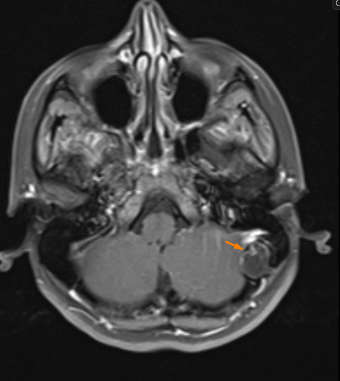
Post-contrast T1 FS Axial Remember: internal enhancement should lead you to consider other diagnoses!
3. DWI: The Identity Card

The lesion glows bright white on DWI (Diffusion Restriction).
DWI: High signal.
ADC: Low signal.
Differential Diagnosis: The Fine Line
It is easy to confuse an epidermoid cyst with arachnoid granulations, hemangiomas, or fibrous dysplasia. However, each has a unique "radiological signature." For instance, why does a hemangioma
behave differently on T1? Or why is a cholesteatoma usually associated with the mastoid antrum?
💡 Academic Pearl: If a bone lesion is lytic on CT, fluid-like on MRI, and shows restricted diffusion on DWI; an Epidermoid Cyst should always be at the top of your list.
📚 Want to Learn More?
You can find detailed differential diagnosis tables, in-depth analysis of enhancement patterns, and the essential Checklist for your reports in my exclusive resources:
👉 [FREE] Join our library to download the Reporting Checklist for this case.
👉 [PREMIUM] Visit our shop for the Extended Case Guide (E-Book /PDF), covering surgical navigation and advanced radiological nuances of Intradiploic Epidermoid Cysts.


Comments