
Epidermoid (Inclusion) Cyst – Multimodal Imaging CaseNo: SST.1.001
Updated: Apr 11


Keywords: Thin capsule , subcutaneous location
🔹 Clinical:
Slowly growing, subcutaneous lesion
Located close to the skin surface
Stable for more than 2 years
🔹 CT findings (non-contrast, 2 years ago):

Central cystic appearance
Surrounding dirty fat planes → chronic inflammatory reaction
Stability over time favors a benign process
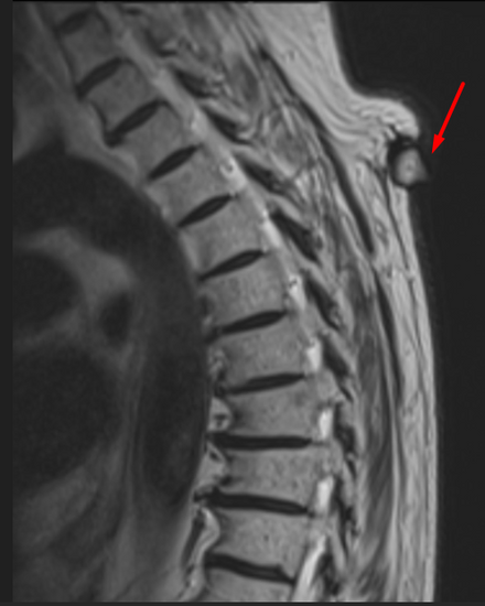
🔹 MRI findings (current):


T1: Hypointense
T2: Mildly hyperintense (not as bright as pure fluid, due to keratin/debris)
Capsule: Thin, well-defined
Base: Sitting on the subcutaneous fat
Protrusion: “Beak/drop-shaped tip” extending from dermis into subcutis
🔹 Why the “beak-shaped protrusion”?

The cyst capsule attaches at the dermis–subcutis interface.
Due to the keratin/debris inside, the cyst tries to expand according to its own volume.
Because of the flexible but limited space between the skin and subcutaneous tissue, as the cyst grows, it narrows downward, forming a pointed tip—appearing “beak/drop-shaped” on MRI.
🔹 Key teaching points:
CT: Dirty fat sign = chronic, benign process
MRI: Beak-shaped protrusion + typical signal intensity
Multimodality + stability strongly support an epidermoid cyst
Differential: lipoma, necrotic lymph node, abscess
---
Comments