


Subependymoma: Differential Diagnosis and Imaging Insights in Older Adults
Clinical Case Presentation
Clinical Case: A 72-year-old female patient presented with non-specific headaches. She has no known history of malignancy, and her neurological examination was entirely normal.
Incidental Findings: Periventricular/subcortical ischemic-gliotic foci, a small calcified meningioma, and a developmental venous anomaly (DVA).
Comment: The headaches are non-specific and unlikely related to the lesion; the finding is most consistent with an incidental subependymoma.
Brain MRI Analysis (T1, T2, FLAIR)
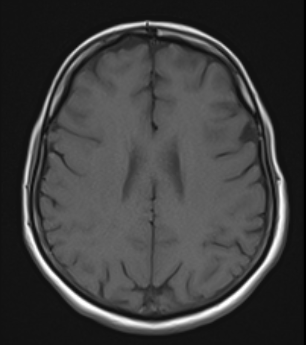
T1WI: The lesion is isointense relative to brain parenchyma. The adjacent ventricular wall is smooth, with no edema in the surrounding parenchyma. Anatomically, it is located in the subependymal region of the left lateral ventricle frontal horn.
T2WI & FLAIR: Isointense on T2 and mildly hyperintense on FLAIR. This finding is consistent with low cellularity and high water content, which is typical for small subependymomas.
Figure 1: Brain MRI Sequences. A 7 mm well-circumscribed nodular lesion is identified in the subependymal region of the frontal horn of the left lateral ventricle (arrow). The lesion is isointense to brain parenchyma on T1W (left) and T2W (middle) images, and mildly hyperintense on FLAIR (right). No surrounding vasogenic edema or mass effect is noted.
Contrast-Enhanced MRI & Advanced Sequences
Post-Contrast T1WI Findings: No contrast enhancement. In the literature, over 80% of subependymomas show no or minimal enhancement due to an intact blood-brain barrier and low vascularity. This case strictly fits that pattern.

Figure 2: Pre- and Post-Contrast T1W MRI. T1-weighted images obtained before (left) and after (right) intravenous contrast administration demonstrate no contrast enhancement within the lesion (arrow). This reflects an intact blood-brain barrier and low vascularity.
Advanced Sequences (DWI & SWI Evaluation):
SWI and DWI sequences were not available in this study.
If SWI were available, we would evaluate for microcalcifications or subtle hemorrhage (lack of blooming would be expected).
On DWI, we would assess for diffusion restriction; given the benign nature, no marked restriction (normal/high ADC) would be expected, excluding high-grade neoplasms.
CT Findings & Differential Diagnosis
CT Findings: Well-demarcated, isodense lesion along the ventricular wall with no gross calcification.
Figure 3: Non-Contrast Brain CT. Axial (left) and sagittal (right) CT scans demonstrate an isoattenuating lesion along the left lateral ventricle wall without evidence of gross calcification or hemorrhage (arrow).
Differential Diagnosis:
Subependymoma: Typical age group, ventricular wall attachment, absence of enhancement, no surrounding edema.
Low-Grade Ependymoma (WHO Grade II): Tends to affect younger patients, exhibits patchy/avid enhancement, and has infiltrative features.
Subependymal Nodular Heterotopia: Isointense to gray matter on ALL pulse sequences, congenital, non-enhancing, and frequently multiple.
Gliomas: Heterogeneous signal intensity with more pronounced contrast enhancement and mass effect.
Calcified Focus / Prior Intraventricular Hemorrhage: Easily differentiated on CT or SWI.
Subependymal Giant Cell Astrocytoma (SEGA): Strongly associated with Tuberous Sclerosis, larger, located near the Foramen of Monro, and shows marked enhancement.
Central Neurocytoma: Typically seen in young adults (20–40 years), attached to the septum pellucidum, highly vascular/heterogeneous with marked enhancement and cystic components.
Summary Trick Points Table
Feature / Point | Explanation & Characteristics |
Origin | Ependymal cells and subependymal glial layer lining the ventricular wall |
Growth | Slow-growing, WHO Grade I, low cell proliferation index |
Vascularization | Low vascularity, no necrosis, intact blood-brain barrier |
Imaging | MRI: T1 isointense, T2 iso/hyperintense, NO contrast enhancement |
Clinical | Mostly asymptomatic; symptoms occur only if CSF flow is obstructed |
Pathology | Clustered glial nuclei embedded in an abundant fibrillary matrix, microcystic changes; GFAP positive, neurofilament negative |
Incidental Finding - Meningioma
Incidental Right Parieto-Occipital Calcified Meningioma. Non-contrast CT (top) demonstrates an 8.5 mm extra-axial calcified nodule at the vertex level in the right posterior parietal region (arrow). Contrast-enhanced T1W MRI (bottom) confirms an incidentally detected calcified meningioma with avid homogeneous enhancement (arrow).
CT Finding: At the vertex level (right posterior parietal region), an extra-axial, 8.5 mm calcified hyperdense nodule is identified.
MRI Finding: Shows marked, homogeneous contrast enhancement on T1+C sequences.
Note: Small meningiomas are frequent incidental findings in older adults, grow slowly, and usually remain asymptomatic.


Incidental Finding – Developmental Venous Anomaly (DVA)
Location: Right posterior parietal region, extending from the periventricular zone toward the vertex and draining into dural sinuses.
CT Finding: Linear/tubular hyperdense venous structures on non-contrast CT.
MRI Finding: Classic "Caput Medusae" appearance on contrast-enhanced MRI.
Note: DVAs are benign extreme variations of normal venous drainage, typically asymptomatic, and require no intervention.

Figure 5: Incidental Right Parietal Developmental Venous Anomaly (DVA). Non-contrast CT (left) demonstrates a linear hyperdense venous structure extending from the periventricular zone toward the vertex (arrow). Contrast-enhanced T1W sagittal MRI (right) exhibits the characteristic "Caput Medusae" appearance draining into dural sinuses (arrow).
Follow-up Plan & Literature Summary
Follow-up Plan:
Baseline SWI sequence addition + follow-up brain MRI every 6–12 months to confirm lesion stability.
Literature demonstrates that small, non-obstructive subependymomas remain stable for years and rarely require surgical resection.
Subependymoma Overview:
Rare, benign (WHO Grade I) intraventricular tumors arising from ependymal/subependymal cells.
Predominantly seen in middle-aged and older adults (50–60s) with a slight male predominance.
Most common locations: Fourth ventricle (~50–60%) and lateral ventricles (~30–40%).
References:
Gaillard F, Sharma R, Walizai T, et al. Subependymoma. Radiopaedia.org.
Jooma R, et al. A Rare Case of Subependymoma with an Atypical Presentation. PMC3223030.
A Subependymoma Clinical Case and Literature Review. PMC9771625.


Comments